Equipment & Capabilities
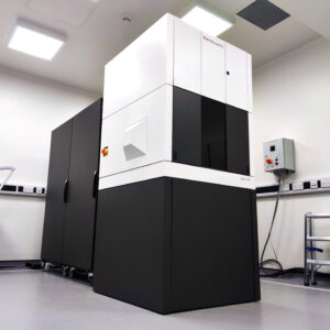 |
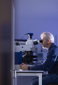
Our scanning/transmission electron microscope enables ultra high-resolution imaging of a wide variety of materials from polymers to metals and ceramics, and also biological materials such as viruses and vesicles. With the included EDS x-ray detector, high-resolution elemental analysis is also possible. The model is a Thermoscientific F200i S/TEM, and it includes highly sensitive Dual-X twin EDS detectors and multi segment S/TEM detector for advanced dark and brightfield imaging. Electron diffraction and electron tomography are also possible. |
 |
Our dual-beam scanning electron microscope is a highly advanced and flexible electron microscope, capable of imaging and analysing a wide variety of materials. Scanning electron microscopy (SEM) is one of the most commonly used methods at the Bridge, especially in combination with EDS. EDS is an x-ray technique that allows the SEM to perform localised elemental analysis.
The instrument we have is a ThermoScientific Scios2 FIB-SEM. It includes a gallium ion column, and semi-automated TEM lemella preparation and slice-and-view 3D imaging. This also includes trinity in-lens detectors and 60mm2 SDD EDX systems. The system includes an easylift micromanipulator, and so can be used to fully prepare electron transparent lamellas for the TEM. |
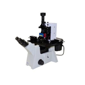
Atomic Force Microscopes (AFMs) are high-resolution microscopes that study the surface texture of samples. Unlike electron microscopes, they can image samples in air, or even in liquid. An AFM forms a three dimensional map of the surface of a sample, and is suitable for a wide range of samples, from biomolecules, to polymers, metals, and nanomaterials. AFM can achieve extremely high contrast on extremely flat samples, such as semiconductor wafers. We have three AFM instruments, described below.
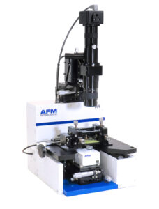 |
AFMWorkshop HR-AFM: A high-resolution complete small-sample AFM with many optional modes, including electrical, magnetic, conducting modes. |
 |
AFMWorkshop
LS-AFM: AFM integrated with brightfield/phase contrast/epifluorescence optical microscopy, and a bio-cell, allowing temperature and environmental control. |
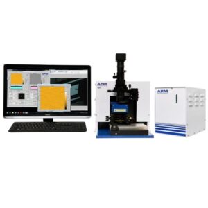 |
AFMWorkshop NP-AFM: Large sample AFM capable of interrogating 20mm wafer-size samples. |
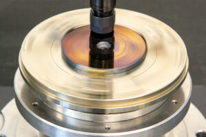 |
The tribometer allows the measurement of friction and wear between any two materials. The instrument allows the measurement of rotating (fixed velocity) movements, or linear (accelerating and decelerating) movement geometries. There are a wide range of sample holders and accessories, allowing the probe to be made of almost any material, and a range of load forces can be used, up to 200 Newtons. Temperature can also be controlled up to 500ºC. A liquid holder, also allows the user to probe the effect of lubrication on the wear and friction. Combination of this instrument with the advanced microscopes in the Bridge allow in-depth study of wear and failure mechanisms.
The instrument we have is an MFT-2000A tribometer from RTEC. |
 |
Nuclear Magnetic Resonance (NMR) spectroscopy is an exquisitely sensitive method for analysing the local magnetic environments of atoms. Within molecules, every atom affects the magnetic environment of other atoms, particularly when they are close together in space or physically bonded to each other.
By monitoring the magnetic environment of the atoms we can determine the structure of molecules, either in isolation or when interacting with other molecules. Since every conceivable environmental condition has a cumulative effect on this magnetic environment – such as molecular interactions, temperature, pH, solution environment – NMR spectroscopy is a very versatile method of analysis. NMR is typically used for structure determination of small molecules, such as chemicals or small biomolecules. NMR can also by used to investigate mixtures of molecules and can even solve 3-dimensional structures of proteins. NMR spectroscopy relies upon exposing the sample of interest to an enormously strong magnetic field, approximately a quarter of a million times as strong as the magnetic field of the Earth, generated by a superconducting magnet. Once exposed to these conditions, each atom within the sample becomes a sensitive barometer of its environment, and will report back on any changes it experiences, such as to temperature, pH, concentration or intra- and inter-molecular interactions. We have the capabilities to routinely analyse a wide variety of elements in a fully automated way, such as hydrogen, carbon, phosphorous and fluorine. Analysis times vary, depending on the sample conditions and analysis desired, but usually take only a few minutes. NMR works well for the analysis of complex mixtures, such as: formulation, identification and quantification of impurities, food forgery, stability studies, metabolomics, reaction monitoring, and elucidating the chemical and three-dimensional structures of compounds and polymers. NMR spectroscopy can be fully quantitative. Our instrument is a Bruker Avance III 500 equipped with CP-MAS H/X (solid probe). |
 |
Raman spectroscopy is a vibrational spectroscopic technique which allows compositional analysis of liquids and solids. The technique can be used to analyse a range of samples (organic and inorganic) to determine structural/compositional changes and for identification of unknown samples.
By coupling Raman spectroscopy to light microscopy, spatial resolution down to 1 micron of compositional changes within a sample can be achieved. In addition, confocal Raman microscopy can be used to perform depth-profiling of a sample giving 3-dimensional chemical resolution. Raman is an identification technique which allows specific chemical bonds in a sample to be determined. Consequently Raman is useful as part of formulation studies or examination of unknown samples to determine the presence of compounds of interest. In addition to determining composition, the technique allows the crystallographic structure of a sample to be determined. This polymorphic information is essential in pharmaceutical studies and can also be used to investigate the crystal structures in mineral and metallurgical samples. By combining the technique with microscopy, spatial chemical resolution can be performed to determine the heterogeneity of a sample. In addition, depth profiling can be carried out to perform 3D studies to look at the penetration of a chemical through a sample e.g. transdermal drug delivery studies. The instrument is a Bruker Senterra II microscope coupled to a RAMII FT Spectrometer, and it is equipped with 534 and 785 nm laser, and also an external fibre-optic probe. |
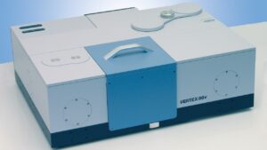 |
The Bruker Vertex 70v spectrometer is a highly sensitive research Fourier-transform infra red (FT-IR) spectrometer. FT-IR spectroscopy is a vibrational spectroscopic technique which allows compositional analysis of liquids, solids and gases.
Molecules absorb infra-red light at wavelengths that correspond to molecular vibrational energies (e.g. bond stretching). The actual energies of light absorbed depends on the strength of the bond and, in turn, on the atoms at either end of the bond and the bond order. Thus, the pattern of light absorbed by a molecule is a composite of all bonds within that molecule and the result is a unique absorption spectrum for each molecule of interest. Other aspects of molecule geometry that impinge on bond strengths and rigidity, such as intra- and inter-molecular interactions, alter the wavelength of light absorbed and hence can be probed by FT-IR. FT-IR is used primarily for the identification of organic compounds due to the readiness of bonds within these molecules to vibrate upon interaction with infra-red radiation. Thus FT-IR is well-suited for the analysis of unknown organic materials such as polymers. By linking FT-IR to thermal analysis (simultaneous thermal analyser (STA)) a multi-pronged approach to sample identification can be performed to increase confidence and reliability. In addition, thermal stability can be assessed by measuring the volatiles released during thermal analysis. The Vertex 70v has wide spectral range (near and mid-infrared ), and a number of sampling accessories. These include ATR, DRIFTS and gas sampling. This last method can be coupled to the STA, to measure the spectra of evolved gases from samples as a function of temperature. |
 |
The dynamic mechanical analyser measures the response of a material to dynamic (oscillating) loads. This enables the user to predict how the material will respond to periodic stresses over a long period of time. Our instrument has accessories to measure the response in tension, compression, 3 point bending and single- or dual-cantilever geometries. The DMA can also be used in thermo mechancial analysis mode to measure coefficient of thermal expansion (CTE). The load applied can be up to 24N and the temperature can be varied up to 500ºC. We also have a humidity generator which enables the study of differnt humidity levels on mechanical properties. Our instrument is a Netzsch DMA 242E Artermis. |
 |
A simultaneous thermal analyser allowing mass change and thermal effects to be measured between -150ºC and 1500ºC. With the STA, we can perform Differential Scanning Calorimetry (DSC) or Thermogravimetric Analysis (TGA). DSC is used to understand phase changes in a material as it is heated. Any exo- or endothermic process can be analysed. TGA measures weight loss with temperature, following thermal degradation process. Since this instrument is coupled to a quadrupole mass spectrometer, we can determine which molecules are released. Our instrument is a Netzsch STA 449, and is coupled to a Netzsch QMS 403D for quadrupole mass spectrometry. |
 |
X-ray diffraction (XRD) is the definitive technique for studying crystalline matter. Diffraction experiments can be done on powders to provide relative quantitation on the levels of different crystalline minerals present, or on single crystal specimens to determine the molecular structure of the crystal at atomic resolution.
At the Bridge we have high-throughput as well as variable temperature and humidity options for powder diffraction to analyse reactions or changes in crystalline forms as a function of environment. A dual source (Cu/Mo) single crystal diffractometer allows for different wavelengths of x-rays to extend the scope of the analyses. This instrument is cryo cooled to increase data quality whilst a plate stage allows for fast screening of crystals from different solution conditions. When X-rays are directed at a sample they are reflected from the electrons of the atoms, if the sample is crystalline, i.e. has long range order, these reflections constructively and destructively interfere with each other giving rise to a specific pattern that is measured. This pattern depends on the internal order of the sample and can be considered like a fingerprint of a crystalline material. Comparison to a database of known patterns helps identify the material in question. Since the X-rays are reflected from the electrons, X-ray diffraction does not give direct evidence on the elements in the material. The analysis of single crystals allows for the determination of the atomic positions in the crystal from the diffraction pattern. This gives rise to accurate molecular elucidation with information on bond lengths and angles in the molecule as well as intermolecular interactions. Our instruments are a Bruker D8 Discover for single crystal diffraction and a Bruker D8 Advance for powder diffraction. |
 |
An advanced materials microscope, which can use variety of contrast methods to observe samples, such as phase contrast, DIC and fluorescence. The microscope is fitted with a rotating sample stage, so is especially suited to polarization microscopy. The system has a highly sensitive Zeiss axiocam camera, using Axiovision software for image acquisition and analysis. With the attached temperature cell, samples may be observed from -150 to +600ºC, as well as allowing observation during DSC experiments. Our microscope is a Zeiss axio Imager.M2m, and is coupled to Linkham DSC600, LNP95 and THMS600 modules. |
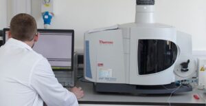 |
Inductively Coupled Plasma Optical Emission Spectroscopy (ICP-OES) is a form of spectroscopy that takes advantage of the fact that the wavelength of the photon emitted by an ion as it recombines with an electron is dependent on the element of the ion. Therefore the photons emitted when a plasma is generated from the analyte can be analysed to reveal the elemental makeup of the analyte. It is fully quantifiable and under favourable circumstances has detection limits in the parts-per-trillion (ppt) range.
Not every element is suitable for analysis by ICP-OES, and certain matrix conditions also make analysis difficult. Therefore care must be taken when preparing an analyte, and to ensure that ICP-OES is an appropriate technique for the desired analysis. The analyte must be fully soluble. In order to ensure this, ICP-OES is often coupled with microwave digestion of the analyte into aqua regia. Under such conditions even the most stubborn analyte can usually be encouraged into solution.
We have two ICP-OES spectrometers, including a top-of-the-range Thermo iCAP 7400. In-line microwave digestion is performed using a Milestone Ethos UP. ICP-OES using this system is fully automated, so that the instrument is capable of high throughput.
|
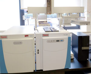 |
Triple quadrupole GC-MS/MS
Gas chromatography combined with triple quadrupole mass spectrometry, known as GC-MS/MS, can operate in three modes: full scan acquisition, selected ion monitoring (SIM), and selective reaction monitoring (SRM). In this system, gas chromatography separates the chemical components of a sample mixture, and mass spectrometry detects, quantifies, and identifies these components. Typical applications of GC-MS/MS include the analysis of volatile and semi-volatile compounds in food, environmental, biological, forensic, and consumer product samples. |
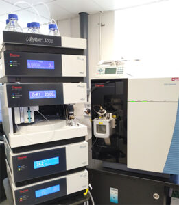 |
Triple quadrupole LC-MS/MS
LC-MS/MS combines high-performance liquid chromatography (HPLC) with triple quadrupole mass spectrometry. The HPLC apparatus separates species in liquid samples, while the mass spectrometer detects, quantifies, and identifies these species, which can be organic or inorganic compounds. The mass spectrometer can operate in full scan mode, selected ion monitoring (SIM) mode, or selective reaction monitoring (SRM) mode. LC-MS/MS is suitable for many applications and is particularly popular for pharmaceutical, biochemical, clinical, and environmental analyses. |
| Ion chromatography (IC) is a type of liquid chromatography that separates ions and ionizable polar molecules in aqueous samples. The charged species are detected and quantified using a conductivity detector. IC is commonly used to analyse common inorganic anions such as F−, H2PO4−, Cl−, Br−, NO3−, and SO42−, as well as cations such as Li+, Na+, NH4+, K+, Mg2+, and Ca2+ in water samples and minerals in geological samples. |
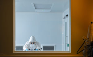 |
The class six clean room is a controlled environment with a guaranteed low level of particulates to prepare and work with materials under ultraclean conditions. |
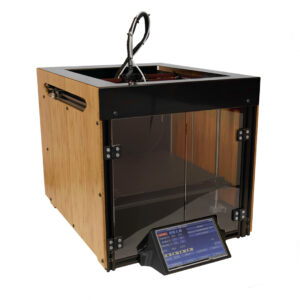 |
The Construct3D Construct 1 is the UK’s fastest 3D printer, boasting exceptional quality, speed, and reliability. With a print speed that is 5x faster than the competition, the Construct 1 is the perfect choice for those who are looking for a high-performance printer that can handle a wide range of printing projects. |
- Henniker Plasma Cleaner – HPT100
- Safematic Sample Coater – CCU-010
- Kambic Environmental Chamber- KK-105 CHLT
- Semi automated sample polishing – Qpol 250
- Precision cut-off saw – Qcut150 A
- Eppendorf centrifuge – 5430